
Translated by AI
CEM Mammography combined with the injection of contrast agents
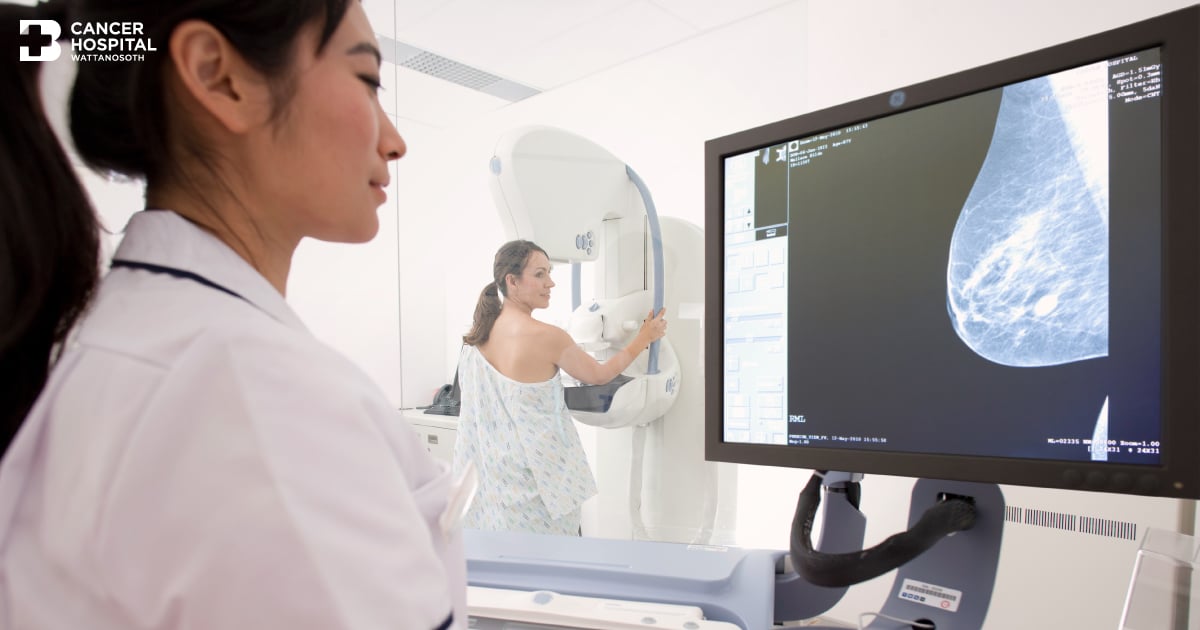
Breast cancer is the most common cancer in women worldwide, including in Thailand. It is considered a silent threat because it often comes without warning signs. Breast examination with a mammogram (Digital Mammography) combined with ultrasound (Ultrasound) remains the primary radiological diagnostic method for breast cancer. However, with advancing technology, additional breast examination methods now provide more detailed diagnostic information, such as Magnetic Resonance Imaging (MRI Breast) and Contrast Enhanced Mammography (CEM).
What is CEM?
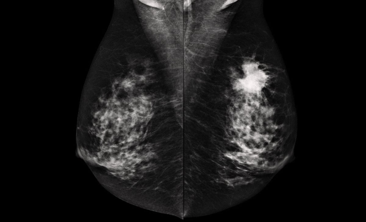
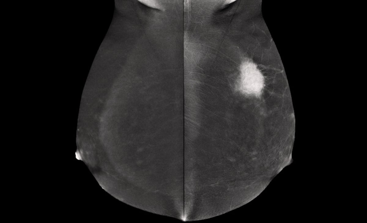
Contrast Enhanced Mammography (CEM) has been approved by the U.S. Food and Drug Administration since 2011 (Food and Drug Administration Approval). Extensive research supports its use, and it is increasingly widespread internationally. It involves injecting contrast media (Iodinated Contrast Media) and using X-rays (Digital Mammography) with a specific dual-energy technique (Dual Energies). The images from both energy levels (Low Energy, High Energy) are processed into Recombined Images. This technique can clearly show suspicious areas in breast tissue, indicating possible abnormal blood supply or breast cancer. CEM is similar to Breast MRI but offers more accuracy compared to standard mammography, especially in patients with dense breast tissue.


Indications for CEM
- Additional screening for breast cancer in asymptomatic women at high risk (Screening for High Risk) such as
- Having a first-degree relative with breast or ovarian cancer, such as a mother or sibling.
- Detection of BRCA1 or BRCA2 genes.
- History of chest radiation between the ages of 10 – 30.
- Additional screening for women with dense breast tissue from previous mammogram results (Supplementation Screening for Women with Dense Breast)
- Diagnosis (Diagnosis)
- Planning treatment before surgery in breast cancer patients (Pre – Surgical Staging of Known Breast Cancer).
- Increasing accuracy in diagnosing breast diseases when mammogram and ultrasound results are inconclusive or complex (Inconclusive Findings on Diagnostic Work Up).
- Monitoring treatment response in breast cancer patients undergoing chemotherapy (Assessment of Tumor Response to Chemotherapy)
- Guiding biopsies for tissue abnormalities detectable only with CEM (CEM Guided Biopsy)
Advantages of CEM
- Improves diagnostic accuracy for detecting breast cancer
- Helps plan surgery in breast cancer patients, reducing repeat surgeries
- Reduces unnecessary breast biopsies
- Saves time and is cheaper than MRI with nearly equal accuracy
- An alternative for patients who cannot undergo MRI
Limitations of CEM
- Higher radiation dose compared to standard mammography but within safety standards.
- Some patients may have allergic reactions to the contrast media.
- Kidney disease patients, those over 60 years old, or those with chronic conditions like diabetes and hypertension should have their kidney function tested (Creatinine Level) within 3 months before the examination.
- Not recommended for patients with silicone breast implants.
- Not recommended for pregnant women unless advised by a physician.
- Breastfeeding patients can undergo the examination with physician approval and can resume breastfeeding 12 – 24 hours after the examination.
Preparation before undergoing CEM
- Provide health history:
- Chronic illnesses, especially asthma, allergies, hyperthyroid, and kidney disease
- Current medications, especially Metformin used for treating diabetes
- Drug and food allergies. Note that seafood allergies are not a contraindication for receiving contrast media
- Previous contrast media exposure
- Side effects from previous examinations if any
- It is recommended to eat light meals, soup, and drink water before and after the examination.
- Avoid applying lotion, powder, perfume, or deodorant on the breast and underarms as they may cause artifacts that affect image interpretation.
- If you have had a previous mammogram, bring the previous results for comparison.
- If you have any breast abnormalities, inform the staff and radiologist performing the examination.
During the CEM procedure
- The nurse will insert an intravenous line in the arm or hand before injecting the contrast media with an automatic injector.
- During the injection, the patient may feel discomfort, warmth, or mild nausea, which usually subsides in a few minutes.
- Within 2 minutes after the injection, the radiologic technologist will position the patient for mammogram imaging using Dual Energies to capture images of both breasts for radiologist interpretation.
Post-examination care
- If the radiologist finds suspicious areas, additional X-ray or ultrasound may be performed, and a biopsy may be considered if abnormalities are detected.
- After the examination, the nurse will remove the IV line, observe the patient for a while, and provide wound care before discharge.
Monitoring for contrast media allergies
If any allergic reactions occur, promptly inform the nurse or treating physician for immediate evaluation:
- Immediate allergic reactions: Rare and usually happen within 1 hour, such as rash, itching, eye irritation, runny nose, etc.
- Severe allergic reactions: Extremely rare, about 0.04%, such as facial or body swelling, difficulty breathing, rapid heartbeat, severe shortness of breath, low blood pressure, or fainting.
Hospitals specializing in breast examination
Bangkok Wattanosoth Cancer Hospital has a breast center equipped with specialized doctors and modern tools. The multidisciplinary team with extensive experience provides efficient breast examination, ensuring accurate results and close care. Early detection of breast cancer increases the chances of complete recovery and reduces the risk of recurrence.
Doctors specializing in breast examination
Dr..Kwansakul Boonsararuxapong, specialist in breast diagnostic imaging, Breast Center, Bangkok Wattanosoth Cancer Hospital
You can click here to make an appointment.
Breast examination package
Breast examination package starts at 5,500 THB